Introduction to the Endoplasmic Reticulum
Endoplasmic reticulum in a cell easy drawing – The endoplasmic reticulum (ER) is a vast, interconnected network of membrane-bound sacs and tubules that extends throughout the cytoplasm of eukaryotic cells. It’s a dynamic organelle crucial for a wide range of cellular processes, acting as both a manufacturing and transport hub. Its intricate structure allows for efficient processing and movement of molecules within the cell.The ER’s extensive network is crucial for its function.
Its proximity to the nucleus and its continuous membrane system facilitates rapid communication and transport of materials between different cellular compartments. This efficient system ensures the smooth and timely execution of many vital cellular tasks.
The Two Types of Endoplasmic Reticulum
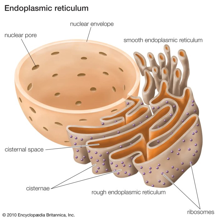
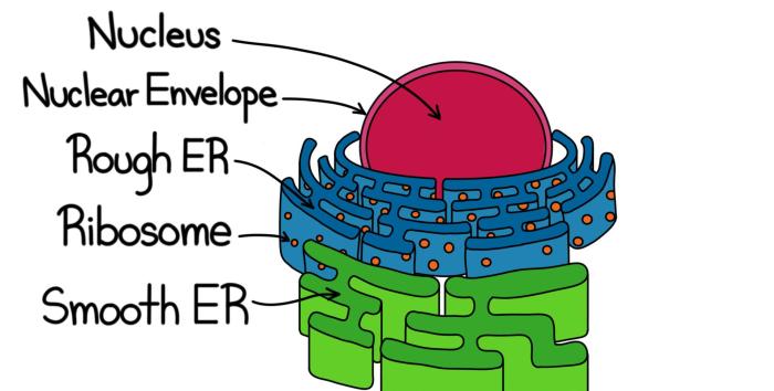
The endoplasmic reticulum is broadly categorized into two distinct types: rough endoplasmic reticulum (RER) and smooth endoplasmic reticulum (SER). These two types, while interconnected, have significantly different structures and functions, reflecting their specialized roles in cellular metabolism.The rough endoplasmic reticulum is studded with ribosomes, giving it its characteristic “rough” appearance under a microscope. These ribosomes are responsible for protein synthesis, specifically those destined for secretion, incorporation into membranes, or transport to other organelles.
The smooth endoplasmic reticulum, lacking ribosomes, plays a key role in lipid synthesis, carbohydrate metabolism, and detoxification processes. Its smooth surface reflects the absence of ribosomes.
Rough Endoplasmic Reticulum: Protein Synthesis and Modification
The RER is the primary site for the synthesis and modification of proteins intended for export from the cell or for use in other organelles. Ribosomes attached to the RER translate messenger RNA (mRNA) into polypeptide chains, which then enter the lumen of the RER for folding and modification. This includes glycosylation (the addition of carbohydrate chains) and disulfide bond formation, crucial steps for proper protein function and stability.
Proteins synthesized in the RER are then packaged into transport vesicles for delivery to the Golgi apparatus for further processing and sorting.
Smooth Endoplasmic Reticulum: Lipid Metabolism and Detoxification
The SER is involved in a diverse range of metabolic processes. Its primary functions include lipid synthesis (including phospholipids and steroids), carbohydrate metabolism, and detoxification of harmful substances. In liver cells, for example, the SER plays a critical role in detoxifying drugs and other toxins. The enzymes within the SER modify these substances, making them more water-soluble and easier to excrete from the body.
The SER also plays a crucial role in calcium ion storage and release, a process essential for muscle contraction and other cellular signaling events.
Analogy for Endoplasmic Reticulum Function
Imagine a large factory with two main departments: one responsible for manufacturing and packaging products (RER) and another for quality control, waste management, and specialized production (SER). The RER receives instructions (mRNA) and assembles proteins (products), folding and modifying them before packaging them for shipment (transport vesicles). The SER, on the other hand, handles various tasks like waste processing (detoxification), managing resources (calcium storage), and producing specific materials (lipids).
The entire factory (ER) is interconnected, ensuring efficient workflow and product delivery.
Easy Drawing of the Endoplasmic Reticulum
A simple diagram of the endoplasmic reticulum (ER) can be surprisingly effective in conveying its key features. While the actual ER is a complex, three-dimensional network, a simplified 2D representation using basic shapes is sufficient for educational purposes, particularly for beginners. This approach focuses on the essential structural components and their spatial relationship within the cell.
Simplified Diagram of the Endoplasmic Reticulum within a Cell
The following describes a basic drawing of a cell containing the endoplasmic reticulum. Imagine a roughly circular cell membrane, represented by a simple oval. Within this oval, the nucleus is represented by a smaller, slightly off-center circle. The ER is depicted as a network of interconnected flattened sacs (cisternae) and tubules, extending throughout the cytoplasm (the area between the nucleus and the cell membrane).
These are represented by parallel lines for the cisternae and slightly curved lines for the tubules, all interconnected to suggest a network. The rough ER, studded with ribosomes (small dots), is differentiated from the smooth ER (lacking ribosomes) by the presence of these dots along the flattened sacs. The overall impression should be one of an extensive, interconnected network filling much of the cell’s interior, but not completely obscuring the nucleus.
Step-by-Step Guide to Drawing the Endoplasmic Reticulum
First, draw a large oval to represent the cell membrane. Then, draw a smaller circle inside the oval to represent the nucleus. Next, draw several parallel lines, slightly curved in places, to represent the cisternae of the endoplasmic reticulum. These lines should extend throughout the cytoplasm, avoiding the nucleus. Add smaller, interconnected, curved lines to represent the tubules of the ER.
Finally, to depict the rough ER, add small dots along some of the parallel lines representing the ribosomes. Remember to leave some areas without ribosomes to represent the smooth ER. The lines representing the ER should be interconnected, showing its network-like structure. The drawing does not need to be perfectly symmetrical; a slightly irregular network better reflects the actual structure.
Key Features of a Basic ER Drawing
A basic drawing of the ER must include several key features to be informative. The most important is the representation of the ER as a network of interconnected sacs (cisternae) and tubules. These should be depicted as interconnected lines, avoiding a disorganized jumble. Another crucial element is the differentiation between rough and smooth ER. This is easily achieved by adding small dots (ribosomes) along some of the cisternae lines.
The relative position of the ER within the cell, extending throughout the cytoplasm but not overlapping the nucleus, should also be clearly illustrated. Finally, the drawing should be simple enough to be easily understood by beginners, yet detailed enough to show the essential characteristics of the ER.
Functions of the Rough Endoplasmic Reticulum: Endoplasmic Reticulum In A Cell Easy Drawing

The rough endoplasmic reticulum (RER), studded with ribosomes, plays a crucial role in protein synthesis and modification within the cell. Its unique structure, characterized by a network of interconnected flattened sacs called cisternae, directly facilitates these essential processes. The intimate association between the RER and ribosomes is key to understanding its multifaceted functions.The rough endoplasmic reticulum’s primary function is the synthesis, folding, modification, and transport of proteins destined for secretion, insertion into cellular membranes, or targeting to other organelles.
Simplifying the complex folds of the endoplasmic reticulum in a cell, for a basic drawing, requires focus on its network-like structure. Think of it as a miniature highway system within the cell, a far cry from the delicate intricacies of, say, an insect’s wing – you can find some great guides for drawing of a insects easy , if that helps you visualize the scale difference.
Returning to our cell, even a simple ER drawing should capture this essential interconnectedness, highlighting its role as a cellular transport hub.
This intricate process relies heavily on the ribosomes embedded in its membrane.
Ribosome Function in Rough ER Protein Synthesis
Ribosomes, the protein synthesis machinery, are the critical components attached to the RER. These ribosomes translate messenger RNA (mRNA) molecules, carrying genetic instructions from the nucleus, into polypeptide chains. The proximity of the ribosomes to the RER membrane is not coincidental; it allows for the newly synthesized proteins to be directly inserted into the RER lumen (interior space) or embedded within the RER membrane itself.
This direct translocation prevents the nascent proteins from entering the cytosol and ensures their proper folding and modification. The RER membrane provides a scaffold for the ribosomes and an environment for protein processing.
Protein Synthesis and its Connection to the Rough ER
Protein synthesis begins in the cytoplasm with the initiation of translation. However, for proteins destined for the RER, a signal sequence – a specific amino acid sequence at the beginning of the polypeptide chain – targets the ribosome-mRNA complex to the RER membrane. The signal sequence interacts with a signal recognition particle (SRP), which temporarily halts translation. The SRP-ribosome-mRNA complex then binds to an SRP receptor on the RER membrane.
Translation resumes, and the growing polypeptide chain is threaded through a protein translocator channel in the RER membrane into the lumen.Once inside the lumen, the protein undergoes a series of modifications, including folding, glycosylation (addition of carbohydrate chains), and disulfide bond formation. These modifications are essential for protein function and stability. Molecular chaperones within the RER lumen assist in proper protein folding, preventing misfolding and aggregation.
Quality control mechanisms ensure only correctly folded proteins are transported further. Incorrectly folded proteins are targeted for degradation.
Examples of Proteins Produced by the Rough ER and Their Functions
Several important proteins are synthesized and processed by the RER. These include:* Secretory proteins: Hormones such as insulin (regulates blood glucose levels) and growth hormone (stimulates growth and cell reproduction), digestive enzymes like amylase (breaks down starch) and pepsin (breaks down proteins), and antibodies (part of the immune system). These proteins are packaged into vesicles for secretion outside the cell.* Membrane proteins: These proteins are integral parts of cellular membranes, including receptors, ion channels, and transporters.
For example, the glucose transporter GLUT4, which facilitates glucose uptake into cells, is synthesized and integrated into the membrane via the RER.* Lysosomal proteins: Lysosomes are organelles responsible for waste degradation. Hydrolytic enzymes, which break down various macromolecules, are synthesized by the RER and targeted to the lysosomes. These enzymes require specific modifications within the RER to function correctly and avoid damaging other cellular components.
Visual Representation

A detailed drawing of the endoplasmic reticulum (ER) necessitates a multi-faceted approach, showcasing not only its intricate structure but also its dynamic interactions with other cellular components. A simple diagram cannot fully capture the complexity of this organelle’s role in protein synthesis, lipid metabolism, and calcium homeostasis. Therefore, a more comprehensive visual representation is crucial for understanding its function within the cell.The following description details a hypothetical, highly detailed drawing of the ER, emphasizing its interconnectedness within the cellular landscape.
Detailed ER Drawing and Description
Imagine a drawing encompassing a significant portion of a eukaryotic cell. The ER is depicted as a network of interconnected, membrane-bound sacs and tubules, extending throughout the cytoplasm. The rough ER (RER), studded with ribosomes (represented as small, dark spheres), is shown in close proximity to the nucleus, its flattened cisternae appearing as stacked pancakes. Ribosomes are actively translating mRNA, with nascent polypeptide chains depicted as thin, string-like structures emerging from the ribosomes and entering the lumen of the RER.
The smooth ER (SER), lacking ribosomes, is portrayed as a network of interconnected tubules, often appearing more tubular and less organized than the RER. It is shown extending from the RER, branching out into the cytoplasm. Transitional elements, where vesicles bud off from the ER, are clearly illustrated, showing the movement of proteins and lipids to other organelles.
The Golgi apparatus, a stack of flattened membrane-bound sacs, is positioned near the RER, with vesicles transporting proteins from the ER to the Golgi for further processing and packaging. Mitochondria, depicted as bean-shaped organelles with inner and outer membranes, are shown in close proximity to the SER, reflecting the SER’s role in lipid metabolism and calcium storage, which are crucial for mitochondrial function.
Finally, the nucleus, with its nuclear envelope continuous with the RER, is included to emphasize the ER’s close association with this central cellular control center. The drawing uses color-coding to distinguish the different components: the RER is a light blue, the SER a light green, ribosomes are dark brown, the Golgi apparatus is a light orange, mitochondria are dark red, and the nucleus is a light purple.
Arrows indicate the flow of proteins and lipids between the different organelles.
Importance of Visualizing ER within the Cell
Visualizing the ER within the context of the entire cell underscores its central role in cellular function. This detailed drawing helps illustrate the ER’s extensive network, highlighting its pervasive influence on protein synthesis, lipid biosynthesis, and calcium signaling. The proximity of the RER to the nucleus emphasizes the direct link between gene expression and protein synthesis. The connections between the SER and mitochondria highlight the interplay between lipid metabolism and energy production.
The representation of vesicles transporting cargo between the ER, Golgi, and other organelles demonstrates the ER’s critical role in the secretory pathway. In essence, a comprehensive visual representation transforms the ER from an abstract concept into a dynamic, interconnected component of a living cell, emphasizing its critical contribution to cellular homeostasis and overall function.
Common Misconceptions about the ER
The endoplasmic reticulum (ER), a vital organelle within eukaryotic cells, is often subject to misunderstandings regarding its structure and function. These misconceptions can stem from simplified diagrams or a lack of appreciation for its dynamic and complex nature. Clarifying these inaccuracies is crucial for a complete understanding of cellular processes.The ER’s intricate network, its diverse roles in protein synthesis and lipid metabolism, and its interactions with other organelles can lead to several common misconceptions.
Addressing these inaccuracies will provide a more accurate and comprehensive picture of this crucial cellular component.
The ER is a Static Structure
A frequent misconception is that the ER is a static, unchanging structure within the cell. In reality, the ER is a highly dynamic organelle constantly undergoing remodeling and reshaping to meet the cell’s fluctuating demands. Its tubules and cisternae are in a state of flux, extending, retracting, and fusing to adapt to changes in protein synthesis, lipid production, and calcium storage needs.
For instance, during periods of increased protein secretion, the rough ER expands to accommodate the higher demand for ribosome binding and protein folding. Conversely, during periods of reduced cellular activity, the ER network can retract and become less extensive. This dynamic nature is essential for the ER’s ability to effectively respond to cellular signals and maintain cellular homeostasis.
The Rough ER Only Handles Protein Synthesis
While the rough ER is heavily involved in protein synthesis, it’s inaccurate to believe that this is its sole function. The rough ER also plays a crucial role in protein folding, modification, and quality control. Newly synthesized proteins undergo extensive processing within the ER lumen, including glycosylation, disulfide bond formation, and chaperone-mediated folding. Proteins that fail to fold correctly are targeted for degradation, preventing the accumulation of misfolded proteins that could disrupt cellular function.
Furthermore, the rough ER contributes to the synthesis of phospholipids and other membrane components, demonstrating its involvement in broader cellular processes beyond solely protein production.
The Smooth ER Exclusively Functions in Lipid Metabolism, Endoplasmic reticulum in a cell easy drawing
Another common misunderstanding is that the smooth ER is solely dedicated to lipid metabolism. Although lipid synthesis is a primary function of the smooth ER, this organelle also plays a significant role in detoxification processes, calcium storage, and carbohydrate metabolism. In liver cells, for example, the smooth ER contains enzymes that metabolize drugs and toxins, protecting the cell from harmful substances.
The smooth ER also acts as a crucial calcium reservoir, regulating calcium ion concentrations within the cytoplasm, a critical process in muscle contraction and signal transduction. Its involvement in carbohydrate metabolism, especially glycogen metabolism in liver and muscle cells, further underscores its multifaceted roles.
FAQ Insights
What are some common mistakes people make when drawing the ER?
Common mistakes include neglecting the interconnectedness of the ER network, failing to differentiate between rough and smooth ER, or drawing it as a single, isolated structure.
How does the ER contribute to cell signaling?
The ER plays a role in calcium signaling, crucial for various cellular processes. It stores and releases calcium ions, influencing many cellular functions.
Can the ER be damaged? What happens if it is?
Yes, the ER can be damaged by various factors, including stress and disease. Damage can lead to disruptions in protein synthesis and other essential cellular functions, potentially resulting in cell death.
How is the ER involved in disease?
ER dysfunction is implicated in several diseases, including various genetic disorders and neurodegenerative conditions. Impaired protein folding and calcium homeostasis are often involved.
